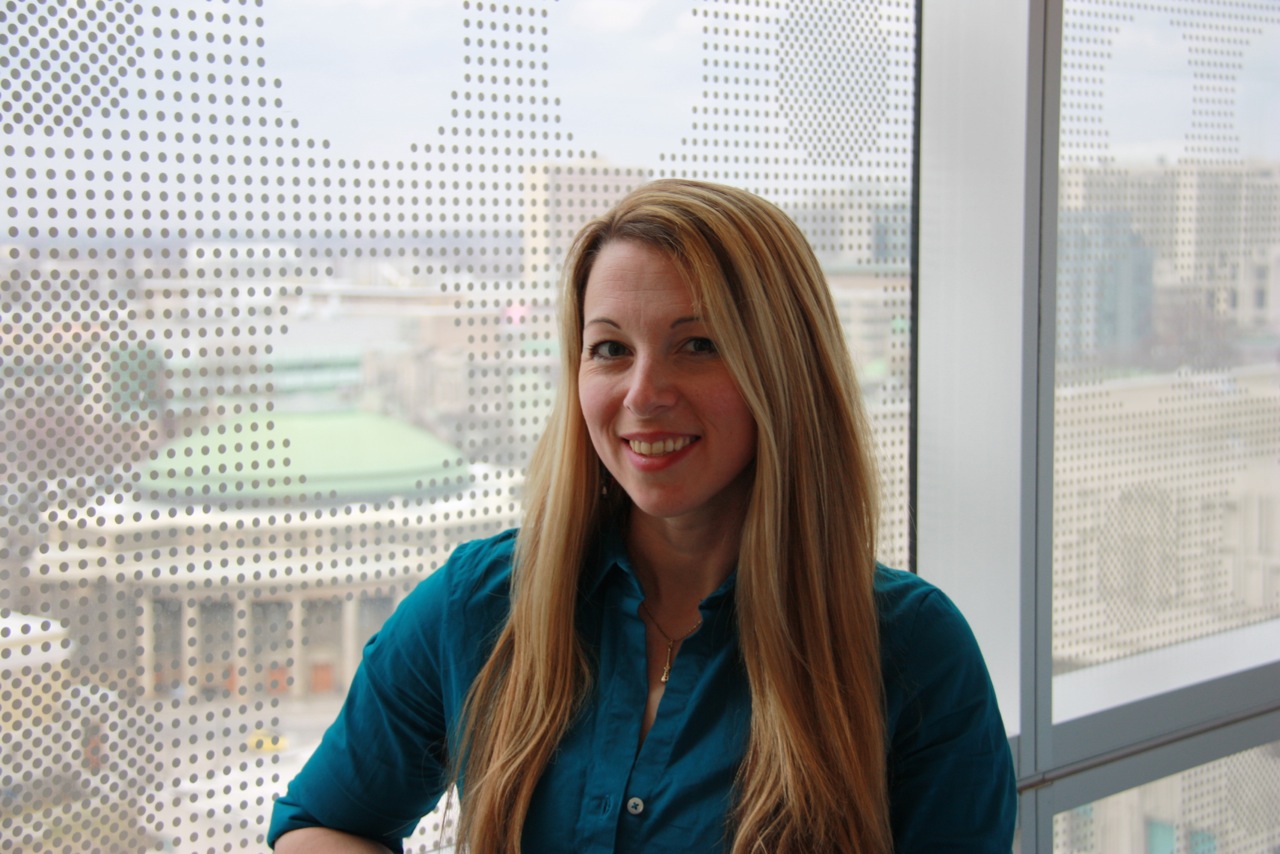
In Profile: Taryn Grieder
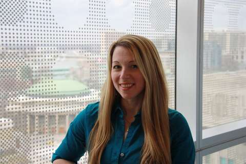
Grieder had been at the University of Toronto since 2000, first as an undergraduate on the Scarborough campus where she studied neuroscience and biology, and later as a doctoral student at the Institute of Medical Science. The Scripps Research Institute in San Diego had offered her a postdoctoral fellowship, but as a new mother with a passion for teaching, she felt the timing and fit weren’t right.
Instead, she enrolled in a Master’s program at the Ontario Institute for Studies in Education.
“It was a good decision, because it made me unique relative to other instructors with a PhD,” says Grieder. “And the program was very relevant, with a focus on higher education, curriculum development, teaching and learning.”
Right after her Master’s degree, Grieder landed an undergraduate sessional lecturer position at U of T teaching synapse neurobiology and the neuroscience of behaviour. Ryerson University also offered her two courses as an instructor, and she kept a staff scientist role with her PhD supervisor, Derek van der Kooy, a Professor in U of T’s Department of Molecular Genetics and the Donnelly Centre for Cellular and Biomolecular Research.
The trifecta let Grieder support herself and her son Luka — and pursue teaching and research. And, she could keep playing sports.
“U of T is a great school for research and education, but it’s also a great place for sports,” says Grieder, who has played intramural hockey, football, volleyball, field hockey, baseball and lacrosse at U of T. She also played varsity lacrosse throughout her doctoral studies and was a first-team Ontario University Athletics all-star in 2013 and 2014. This past year she captained the Blues to a provincial bronze medal.
Grieder’s Toronto ties have also kept her close to her family — a key influence on her athletic and academic interests. She credits her dad for sparking her interest in baseball at the age of five, and she chose to study neuroscience as an undergrad after her grandmother passed away with Alzheimer’s disease. As a graduate researcher she focused on nicotine addiction, in part because all five of her immediate family members were smokers.
Last year, Grieder and her colleagues at Scripps published a paper in Nature Neuroscience that opened a new line of research into why people smoke. The paper showed the brain’s stress and reward systems are linked during nicotine addiction and withdrawal through a neurotransmitter called corticotropin-releasing factor, or CRF. The finding upended conventional thinking about the independent function of the stress and reward systems.
“Taryn is eager to problem-solve in the lab and to explore unusual hypotheses. She has excellent technical skills and seems to have a green thumb in carrying out experiments,” says van der Kooy, who has overseen Grieder’s lab work for 10 years.
Grieder is now studying other molecular receptors that may link the brain’s stress and reward systems, and she wants to continue lab work at least part-time. “It’s also great to be able to discuss research with my students,” she says.
Mandy Yee will finish her undergraduate degree at U of T this spring. She took the two courses Grieder teaches and says she appreciated hearing about Grieder’s addiction research in class, along with other timely studies on neuroscience and cell biology.
Yee also says Grieder is a great mentor.
“Dr. Grieder was the most approachable professor I had as an undergraduate. Her lectures were well prepared, pitched at the right level and very engaging. And she gave me great advice about research and applying to graduate school,” says Yee, who hopes to start a Master’s program at the Institute of Medical Science this fall.
Grieder wants to stay at U of T, but she finds being a sessional lecturer and contract research scientist stressful. She has applied for a full-time lecturer position at U of T and expects to hear the results in June.
And she’s taking a run at another extracurricular activity: “This time next year I’ll be happy if I’m coaching my beloved Varsity Blues women’s lacrosse team — and of course doing research and teaching,” she says.

Optimize this page for search engines by customizing the Meta Title and Meta Description fields.
Use the Google Search Result Preview Tool to test different content ideas.
Select a Meta Image to tell a social media platform what image to use when sharing.
If blank, different social platforms like LinkedIn will randomly select an image on the page to appear on shared posts.
Posts with images generally perform better on social media so it is worth selecting an engaging image.
By Jim Oldfield
Study May Identify New Cause of Brain Bleeds in Fetuses and Newborns
A newly discovered bodily process in mice may explain why some human fetuses who have different antigens than their mothers suffer life-threatening brain bleeds, according to a new study.

“Antigens are like the body’s national flag. They’re planted on each cell in the body and tell the immune system whether something in the body, such as a bacteria or virus, is foreign,” said Heyu Ni, a Professor in the Departments of Laboratory Medicine and Pathobiology, Medicine and Physiology. “Because each parent’s DNA is different, a fetus can have different antigens, or flags, on some cells than his or her mom. When a mom’s immune system identifies those cells as foreign to the body it attacks them, which can cause brain bleeds and result in neurological impairment or even death.”
The condition where mothers and fetuses have different antigens is called fetal and neonatal alloimmune thrombocytopenia, or FNAIT. It affects about one in 1,000 live births. Fetuses experience bleeding in the brain in about 10 to 20 per cent of FNAIT cases. The disease can also cause miscarriages, although that has not been well studied.
Platelets—cells in the blood that help form blood clots and stop bleeding—are one of the cell types that commonly have different antigens. Because they are often different in the mother and fetus, they can be targeted by the mother’s immune system. Until now, low amounts of platelets were considered the cause of brain bleeds in fetuses and newborns.
“Our research challenges the idea that low platelet counts are responsible for fetal brain bleeds and instead shows that the immune system’s attack on the new blood vessel cells in the brain are more likely responsible,” said Ni, who is a scientist in the Keenan Research Centre for Biomedical Science of St. Michael’s Hospital as well a Canadian Blood Services scientist. “An antigen, called beta 3 integrin, is found both on platelets and on the cells responsible for developing blood vessel in fetuses.”
Newborn platelet levels are tested at birth since it’s believed a lower platelet count signifies the newborn lacks the ability to stop bleeding. A safe level of platelets for newborns is between 150 million and 450 million cells per ml of blood. Babies with low platelet counts (less than 150 million cells per ml of blood) are usually treated right away with platelet transfusions.
“What we’ve discovered means that platelet transfusions are necessary to control bleeding after birth but may not be an effective therapy for brain bleeds in fetuses since platelets may be not essential to stop fetal bleeding,” said Ni. “We should consider different therapies to prevent brain bleeds and ensure blood vessels in the brain are developed properly before birth.”
Ni’s research team also looked at the potential treatment of intravenous immunoglobulin transfusions. He said IVIG—made of plasma from donated blood—may be an effective therapy to control this devastating disease, although more research is needed to confirm this.
The study was published in The Journal of Clinical Investigation. The project was supported by Canadian Institutes of Health Research, the Canadian Foundation for Innovation and Canadian Blood Services.
A newly discovered bodily process in mice may explain why some human fetuses who have different antigens than their mothers suffer life-threatening brain bleeds, according to a new study.
“Antigens are like the body’s national flag. They’re planted on each cell in the body and tell the immune system whether something in the body, such as a bacteria or virus, is foreign,” said Heyu Ni, a Professor in the Departments of Laboratory Medicine and Pathobiology, Medicine and Physiology. “Because each parent’s DNA is different, a fetus can have different antigens, or flags, on some cells than his or her mom. When a mom’s immune system identifies those cells as foreign to the body it attacks them, which can cause brain bleeds and result in neurological impairment or even death.”
The condition where mothers and fetuses have different antigens is called fetal and neonatal alloimmune thrombocytopenia, or FNAIT. It affects about one in 1,000 live births. Fetuses experience bleeding in the brain in about 10 to 20 per cent of FNAIT cases. The disease can also cause miscarriages, although that has not been well studied.
Platelets—cells in the blood that help form blood clots and stop bleeding—are one of the cell types that commonly have different antigens. Because they are often different in the mother and fetus, they can be targeted by the mother’s immune system. Until now, low amounts of platelets were considered the cause of brain bleeds in fetuses and newborns.
“Our research challenges the idea that low platelet counts are responsible for fetal brain bleeds and instead shows that the immune system’s attack on the new blood vessel cells in the brain are more likely responsible,” said Ni, who is a scientist in the Keenan Research Centre for Biomedical Science of St. Michael’s Hospital as well a Canadian Blood Services scientist. “An antigen, called beta 3 integrin, is found both on platelets and on the cells responsible for developing blood vessel in fetuses.”
Newborn platelet levels are tested at birth since it’s believed a lower platelet count signifies the newborn lacks the ability to stop bleeding. A safe level of platelets for newborns is between 150 million and 450 million cells per ml of blood. Babies with low platelet counts (less than 150 million cells per ml of blood) are usually treated right away with platelet transfusions.
“What we’ve discovered means that platelet transfusions are necessary to control bleeding after birth but may not be an effective therapy for brain bleeds in fetuses since platelets may be not essential to stop fetal bleeding,” said Ni. “We should consider different therapies to prevent brain bleeds and ensure blood vessels in the brain are developed properly before birth.”
Ni’s research team also looked at the potential treatment of intravenous immunoglobulin transfusions. He said IVIG—made of plasma from donated blood—may be an effective therapy to control this devastating disease, although more research is needed to confirm this.
The study was published in The Journal of Clinical Investigation. The project was supported by Canadian Institutes of Health Research, the Canadian Foundation for Innovation and Canadian Blood Services.
Optimize this page for search engines by customizing the Meta Title and Meta Description fields.
Use the Google Search Result Preview Tool to test different content ideas.
Select a Meta Image to tell a social media platform what image to use when sharing.
If blank, different social platforms like LinkedIn will randomly select an image on the page to appear on shared posts.
Posts with images generally perform better on social media so it is worth selecting an engaging image.
Melissa Di Costanzo
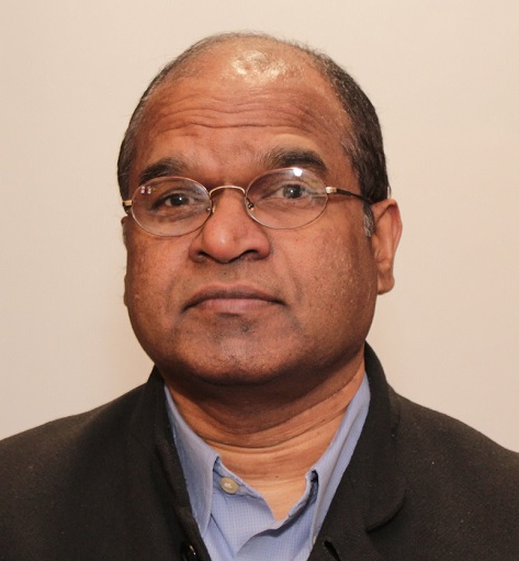
Solving Inflammation at the Source: Q&A with Professor Nades Palaniyar

How does the body’s immune system react to invading microorganisms and how can that normally protective function lead to inflammation and disease? Researchers from the Department of Laboratory Medicine and Pathobiology at U of T believe they’ve found a missing link in understanding this unusual connection.
Professor Nades Palaniyar, along with PhD student David Douda and Postdoctoral Fellow Meraj Khan, has revealed a new pathway in an immune response called NETosis.
When bacteria invade, the human body reacts within minutes and sends out neutrophils—a type of white blood cell that engulfs and digests microorganisms. But now researchers know that neutrophils can also cast nets, called neutrophil extracellular traps (NETs), that ensnare microbes to control infection.
While this process is essential for the immune system to react to acute infections, it can also cause severe long-term damage. When neutrophils cast nets, they release toxins that can cause excess inflammation and damage to surrounding tissues and organs. This inflammation can lead to or worsen debilitating disorders including lupus, rheumatoid arthritis, cystic fibrosis, difficult-to-treat neutrophilic asthma and several vascular diseases.
In this Q&A, Palaniyar discusses his findings recently published in the Proceedings of the National Academy of Sciences (PNAS). In an area with few treatment options, his work provides hope for patients with inflammatory and autoimmune diseases.
What did you propose in your recent PNAS paper?
Two major types of NETosis have been described to date, but researchers didn’t understand how or why they operated differently. One type depends on an enzyme called NADPH oxidase 2 (NOX2). The pathways that depend on this enzyme are reasonably well-established.
We’ve identified the mechanistic details of the second type of NETosis that doesn’t depend on the NOX2 enzyme. In this paper, we show that a calcium-activated potassium channel (SK3) mitochondrial reactive oxygen species (ROS) and specific enzymes regulate this pathway.
We’re hoping to target different components of NETosis pathways to identify better drugs. We’re currently screening thousands of existing FDA-approved drugs to identify effective treatment options. We’re hoping to find a drug that will eliminate the chronic inflammation caused by NETosis, while still allowing the immune system to deal with an acute infection.
How could your discovery change how patients are treated?
Currently, we don’t have many options for dealing with inflammatory or autoimmune diseases. Clinicians treat most of these patients with steroids to suppress their immune systems, or they use other non-steroid drugs such as aspirin and ibuprofen.
These drugs have considerable side effects. For example, using steroids on patients who are already immunosuppressed can lead to other infections. Steroids also have other side effects because they regulate many pathways, and they don’t always work on all patients.
Once we know the molecular mechanisms that regulate chronic inflammation, it is possible to identify a potent drug with minimal side effects. We’re hoping that our discovery will lead to better options for patients.
What inspired you to conduct research in this area?
I always like to investigate new topics. About ten years ago, researchers identified NETosis, and determined that NETs were made of DNA and anti-microbial proteins. During my postdoctoral fellowship at Oxford University, I published a paper based on these findings. When I moved to Toronto and joined SickKids as a scientist and LMP as an assistant professor, I continued my work and I’ve been studying NETosis ever since.
We were right there at the beginning, and there were no models even to study NETs in vivo. We’ve devised many in vivo-ex vivo models and tests to study NETosis, so I really feel that we’re inventors discovering something entirely new.
Sometimes, when I open my eyes at the end of my meditation, I suddenly see answers to NET questions. I even wake up in the middle of the night and write equations and new pathways! I have a file full of these outlines and ideas. I talk about these new ideas for hours with my trainees, who are fascinated by NETs. The elegance of the immune system inspires me to do more and more.
What are your next steps?
Now that we know certain players within the cells, we’re currently trying to identify FDA-approved drugs and new compounds that will target specific pathways. These drugs should stop NET formation while maintaining the immune function of the cells. We already have excellent results from our first drug screen. They were exactly the groups of drugs that I had predicted would work. The whole process of identifying the best compounds will take close to two years.
If we identify a compound that we think will target one or both of these pathways, we’ll then move to in vivo studies. I’m excited about advancing this research and understanding how to block the pathways. I collaborate well with many clinicians. Dr Harmut Grasemann, who is a co-author in this study, is an outstanding collaborator for CF lung diseases I think it is very important for scientists and clinicians to interact efficiently to bring discoveries to the clinic at the earliest possible time.
What kind of technology are you using to find suitable treatments?
We have an elaborate robotic system at SickKids that uses echo vibrations to deposit tiny amounts of drugs very precisely. It’s so sensitive and efficient that we can test thousands of drugs from these libraries at the same time—one screen will test 1,280 compounds. We’ll first test 4,000 FDA-approved drugs, and then test the library with 40,000 new compounds.
We’re also using another new technology at SickKids that combines flow cytometry and mass spectrometry, called CyToF, to identify other enzymes that might be involved in NETosis. This approach can generate 36-dimensonal precise information. The ordinary human mind cannot process information beyond a few dimensions.
LMP alumnus David Douda worked on this research. What was his role?
Students who are really open minded and want to explore big ideas thrive in my lab. David was one of those students, and in 2011 he helped to establish the first in vivo model to study NETs in the lungs. While in the lab he published ten papers as a primary or a co-author and four more will be published soon. After he completed his PhD in 2013, he moved to Brigham and Women’s Hospital, Harvard Medical School for his postdoctoral training. Meraj did a lot of experiments that were necessary to complete the work; therefore, both of them share the first authorship. Meraj is now taking the work to the next level.
I expect, and encourage, everyone in my lab to do more than one project and David started this PNAS work as a side project. It turned out to be great. I have to find a few more PhD students like David and postdocs like Meraj to churn out much more new information in this hot field.
I give my trainees new project ideas to explore, and ask them to find a project that they are passionate about. They have to fall in love what they’re doing. When you receive a PhD degree, you are getting a license that says to the world that you are an independent thinker or a philosopher. You need to learn to think big, and to think on your own. If you do what you love, you will love what you do.
Optimize this page for search engines by customizing the Meta Title and Meta Description fields.
Use the Google Search Result Preview Tool to test different content ideas.
Select a Meta Image to tell a social media platform what image to use when sharing.
If blank, different social platforms like LinkedIn will randomly select an image on the page to appear on shared posts.
Posts with images generally perform better on social media so it is worth selecting an engaging image.
Katie Babcock
Media and Mental Health
To Bruce Ballon, the Internet is like an ocean of information, communication and simulation. To help people navigate these potentially dangerous waters, he is throwing out a life ring.

While smart phones, social media and online gaming become more integrated into our lives, Ballon says it is important for people to reflect on their own reliance on devices and apps. To help with that process, Ballon wrote the free e-book SWIMMING IN CYBER: Learning to live HEALTHILY in the intersections of the virtual and real worlds.
Ballon is an Associate Professor in the Department of Psychiatry and Head of Advanced Clinical and Educational Services for Problem Gambling, Gaming and Internet Addiction at the Centre for Addiction and Mental Health (CAMH). Many of the people he sees in his clinical practice, which supports young people and people with addictions, have co-occurring issues like depression, anxiety or Autism Spectrum Disorders. They use technology to try to deal with their condition just like some people self-medicate with drugs.
“For example, if someone has social anxiety, they may feel more comfortable online than they do in real life. They may say, ‘I can feel very powerful here in this virtual world. In the real world, I feel very disempowered. Which one is more valuable to me? Well, I like the virtual one, even though it’s causing me problems in the long run, it serves a purpose,’” Ballon says. “And so, even though we’re dealing with the underlying or concurrent mental health issue, we still have to deal with the video game or the Internet or else it will just keep on going.”
Computers are everywhere — in schools, at work and in many people’s homes — so simply ignoring them is not an option. Ballon is helping patients find strategies for using technology while protecting themselves from allowing it to drain all of their time or money.
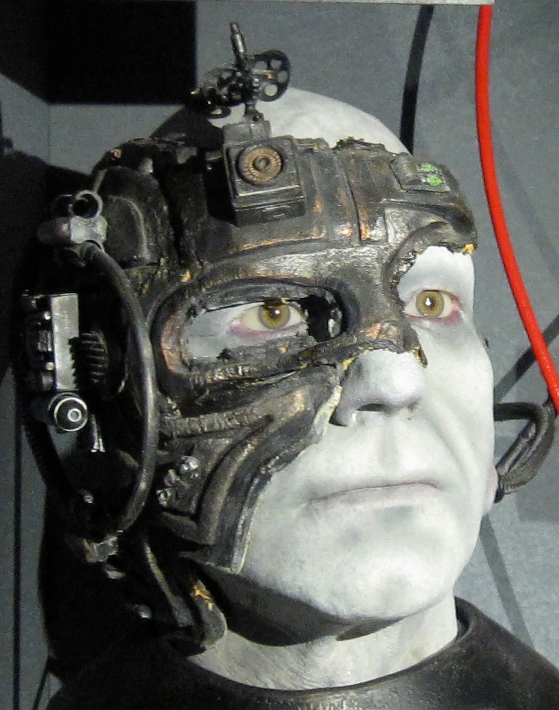
“Technology is now pretty much totally overlapped with our reality,” says Ballon, who is also the Director of Education at the Simulation Ontario Network of Excellence (SIM-one). “Everyone walks around with cell phones, looking like creatures from Star Trek called The Borg, which are cyborgs that are networked together. In my clinical practice, I see people with addictive behaviors like over-using social media connections, online pornography, gambling or video games. I wrote the book to start a conversation about how to use technology in a healthy way.”
Technology and media can also influence us in other ways, says Ballon, who also points to people’s use of brand names as verbs, such as Googling. He explains most people wouldn’t say they “internet search” to find information. Instead, they Google.
“We often don’t even realize it is happening,” says Ballon. “Sometimes when I teach, I ask people what they imagine when I say the word, ‘addict’. Usually, it’s somebody shooting up in an alleyway with needles. As an addiction doctor, I know that is a very small percentage of the addiction field. So why do we think that? Because of what’s portrayed in the media. We need to be more self-reflective to understand the messages coming in and what they mean for us.”
Ballon’s book can be downloaded from Amazon, iBooks, Smashwords and Barnes & Noble.
Click here to read more about Ballon’s work.
To Bruce Ballon, the Internet is like an ocean of information, communication and simulation. To help people navigate these potentially dangerous waters, he is throwing out a life ring.
While smart phones, social media and online gaming become more integrated into our lives, Ballon says it is important for people to reflect on their own reliance on devices and apps. To help with that process, Ballon wrote the free e-book SWIMMING IN CYBER: Learning to live HEALTHILY in the intersections of the virtual and real worlds.
Ballon is an Associate Professor in the Department of Psychiatry and Head of Advanced Clinical and Educational Services for Problem Gambling, Gaming and Internet Addiction at the Centre for Addiction and Mental Health (CAMH). Many of the people he sees in his clinical practice, which supports young people and people with addictions, have co-occurring issues like depression, anxiety or Autism Spectrum Disorders. They use technology to try to deal with their condition just like some people self-medicate with drugs.
“For example, if someone has social anxiety, they may feel more comfortable online than they do in real life. They may say, ‘I can feel very powerful here in this virtual world. In the real world, I feel very disempowered. Which one is more valuable to me? Well, I like the virtual one, even though it’s causing me problems in the long run, it serves a purpose,’” Ballon says. “And so, even though we’re dealing with the underlying or concurrent mental health issue, we still have to deal with the video game or the Internet or else it will just keep on going.”
Computers are everywhere — in schools, at work and in many people’s homes — so simply ignoring them is not an option. Ballon is helping patients find strategies for using technology while protecting themselves from allowing it to drain all of their time or money.
“Technology is now pretty much totally overlapped with our reality,” says Ballon, who is also the Director of Education at the Simulation Ontario Network of Excellence (SIM-one). “Everyone walks around with cell phones, looking like creatures from Star Trek called The Borg, which are cyborgs that are networked together. In my clinical practice, I see people with addictive behaviors like over-using social media connections, online pornography, gambling or video games. I wrote the book to start a conversation about how to use technology in a healthy way.”
Technology and media can also influence us in other ways, says Ballon, who also points to people’s use of brand names as verbs, such as Googling. He explains most people wouldn’t say they “internet search” to find information. Instead, they Google.
“We often don’t even realize it is happening,” says Ballon. “Sometimes when I teach, I ask people what they imagine when I say the word, ‘addict’. Usually, it’s somebody shooting up in an alleyway with needles. As an addiction doctor, I know that is a very small percentage of the addiction field. So why do we think that? Because of what’s portrayed in the media. We need to be more self-reflective to understand the messages coming in and what they mean for us.”
Ballon’s book can be downloaded from Amazon, iBooks, Smashwords and Barnes & Noble.
Click here to read more about Ballon’s work.
Optimize this page for search engines by customizing the Meta Title and Meta Description fields.
Use the Google Search Result Preview Tool to test different content ideas.
Select a Meta Image to tell a social media platform what image to use when sharing.
If blank, different social platforms like LinkedIn will randomly select an image on the page to appear on shared posts.
Posts with images generally perform better on social media so it is worth selecting an engaging image.
Erin Howe
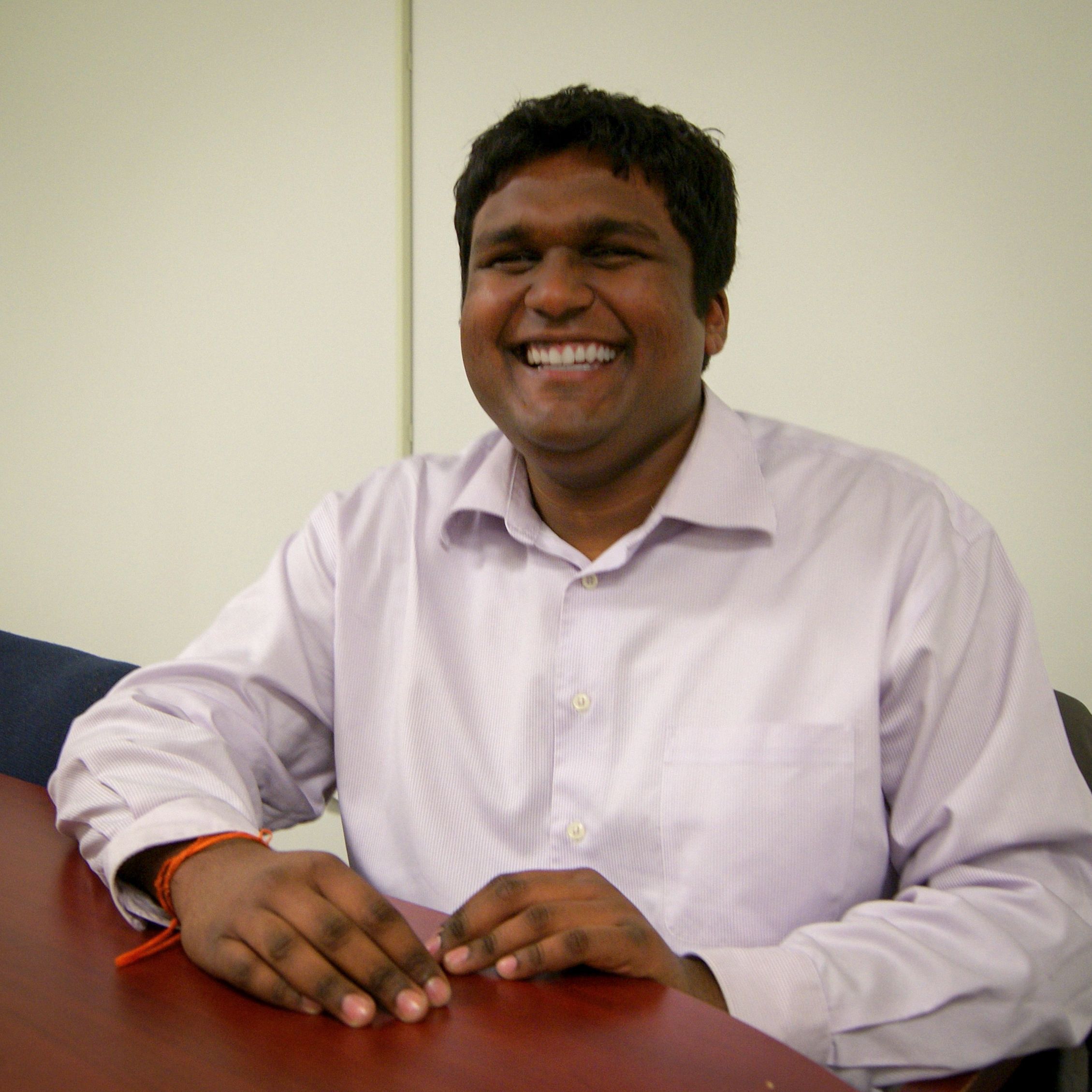
Faces of U of T Medicine: Narayan Chattergoon

Medical students have to be professional multitaskers, but Narayan Chattergoon takes it to a whole new level. Chattergoon is President of U of T Medicine’s Medical Society, a student group that represents all undergraduate medical students. He spoke to writer Suniya Kukaswadia about what it’s like to be the student leader at one of Canada’s top medical schools.
Name: Narayan Chattergoon
Program/year: Second-year MD student
Role/position: President, Medical Society (MedSoc)
Tell us about your work with MedSoc.
As President, I represent all undergraduate medical students. I oversee MedSoc’s portfolios, help set priorities for the Society and represent the Faculty at health conferences. I regularly meet with the Faculty’s senior leadership to discuss any concerns or ideas students have.
What inspired you to get involved with MedSoc?
I’m what I call a “human switchboard” — I like being the go-to person for people who need help. My fellow students come to me if they need to contact a faculty member or need help with a problem. The MedSoc President role really spoke to me because it’s all about connecting with people and representing the interests of medical students.
What do you find most exciting about your role as President?
The best part of the job is working with all the amazing people. The student body is so diverse and engaged, and faculty members are really committed to giving us a positive experience. It’s incredibly rewarding to work together and come up with solutions that have positive impacts and take everyone into account.
What do you hope to accomplish?
I want to work with students and staff to spread the word about all the great things happening at the Faculty. Everyone should have access to the services they need. I want to be accessible, accountable, helpful, and a strong advocate for students.
Kim Blakely, MedSoc’s previous President was working towards greater accountability, transparency and connectivity with students. She also set the wheels in motion for Med Soc to become a registered not-for-profit corporation. I want to build on all that.
How do you think your work with MedSoc will impact your career as a physician?
Physicians need to have strong communication skills. They often have to work with others to make serious decisions with competing priorities and limited resources. It’s important to be creative, to ensure everyone knows that they are valued and come up with solutions together. I’m developing these vital skills through my experience in Med Soc. In the future, I may end up in a managerial or administrative position, and my experience with the students and faculty will be a great foundation.
What's your favourite thing about the Faculty of Medicine?
I’m able to tap into and learn from people who have a wealth of unique experiences. The faculty, staff and students are some of the most caring, dedicated and committed people that I know. This is one of the most inspiring environments I could be in. I feel privileged to be a MD student here, and I love coming to school every day.
Faces of U of T Medicine introduces you to some of the interesting men and women studying in the Faculty of Medicine. From advising political leaders to providing care to Toronto’s most vulnerable populations, our students are making an impact on communities at home and around the world.
Do you have an interesting story to share? Contact Suniya Kukaswadia at Suniya.Kukaswadia@utoronto.ca.
Photo by Paige Zhang
Optimize this page for search engines by customizing the Meta Title and Meta Description fields.
Use the Google Search Result Preview Tool to test different content ideas.
Select a Meta Image to tell a social media platform what image to use when sharing.
If blank, different social platforms like LinkedIn will randomly select an image on the page to appear on shared posts.
Posts with images generally perform better on social media so it is worth selecting an engaging image.
Suniya Kukaswadia
Interprofessional Education Ensures Healthcare is Stronger

Differing perspectives make experiences richer for health care professionals and the people they help.
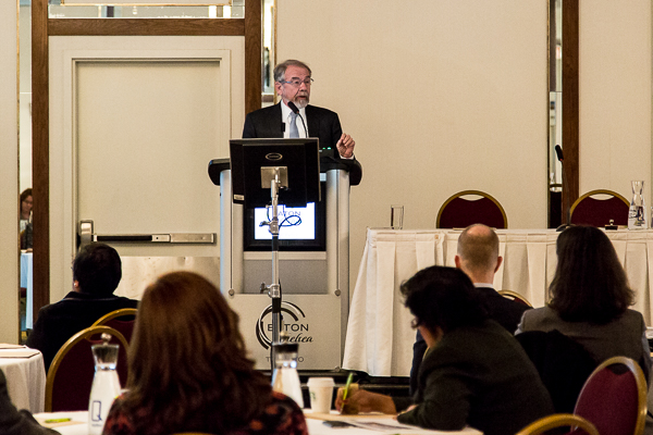
That was one of the messages delivered by George Thibault, the keynote speaker at Reaching the Summit:Leading the Way From Interprofessional Education to Practice. Thibault is the President of the Josiah Macy Jr. Foundation, which works to improve American health care and supports projects that broadens and improves health profession education.
The conference took place December 2 at the Eaton Chelsea Hotel. More than 100 researchers, educators, clinicians and hospital leaders took part in the event, which featured discussions about best practices in Interprofessional Education (IPE) and Interprofessional Practice (IPP).
“We need to partner with each other as health professionals, but we also need to work with the patients and communities we serve,” said Thibault. “IPE is not an end in itself. We should be doing it because the goal is to improve patient care.”
Chrystal Gomes, a patient advocate and stand-up comedienne provided her perspective on navigating the health care system. She lives with numerous ailments including Multiple Sclerosis, depression and chronic pain. She is also a caregiver to her mother who lives with Alzheimer’s Disease. Her experiences highlight the need for collaborative, patient-centered care.
Gomes spoke about the importance of empowering professionals to work together and communicate with their patients. She also expressed a need for open-mindedness to become a sought-after trait in prospective health professionals.
“IPE is a common theme across health professions,” said Sarita Verma, Deputy Dean of the Faculty of Medicine at the University of Toronto. “This model of collaborative learning is tremendously beneficial for students and patients alike. The impact on the health system has included reduced wait times, better coordinated care, and more efficient work patterns.”
Dr. Bob Bell, Ontario’s Deputy Minister of Health and Long-Term Care was another special guest at the event and delivered the opening remarks. Leaders in IPE also led discussions and breakout sessions aimed at increasing collaborative learning in the future, evaluating the success of IPE and nurturing faculty development.
“We know we have a great Toronto Model, we have superb integration across the health sciences, we have the breadth and depth of unparalleled resources here,” said Verma. “We’ve got lots of ideas about Canadian experiences in IPE, and it’s time to take these to the next level.”
Read a Q&A with Dr. George Thibault as well as more on IPE in the Fall 2014 issue of U of T Medicine magazine.
Differing perspectives make experiences richer for health care professionals and the people they help.
That was one of the messages delivered by George Thibault, the keynote speaker at Reaching the Summit:Leading the Way From Interprofessional Education to Practice. Thibault is the President of the Josiah Macy Jr. Foundation, which works to improve American health care and supports projects that broadens and improves health profession education.
The conference took place December 2 at the Eaton Chelsea Hotel. More than 100 researchers, educators, clinicians and hospital leaders took part in the event, which featured discussions about best practices in Interprofessional Education (IPE) and Interprofessional Practice (IPP).
“We need to partner with each other as health professionals, but we also need to work with the patients and communities we serve,” said Thibault. “IPE is not an end in itself. We should be doing it because the goal is to improve patient care.”
Chrystal Gomes, a patient advocate and stand-up comedienne provided her perspective on navigating the health care system. She lives with numerous ailments including Multiple Sclerosis, depression and chronic pain. She is also a caregiver to her mother who lives with Alzheimer’s Disease. Her experiences highlight the need for collaborative, patient-centered care.
Gomes spoke about the importance of empowering professionals to work together and communicate with their patients. She also expressed a need for open-mindedness to become a sought-after trait in prospective health professionals.
“IPE is a common theme across health professions,” said Sarita Verma, Deputy Dean of the Faculty of Medicine at the University of Toronto. “This model of collaborative learning is tremendously beneficial for students and patients alike. The impact on the health system has included reduced wait times, better coordinated care, and more efficient work patterns.”
Dr. Bob Bell, Ontario’s Deputy Minister of Health and Long-Term Care was another special guest at the event and delivered the opening remarks. Leaders in IPE also led discussions and breakout sessions aimed at increasing collaborative learning in the future, evaluating the success of IPE and nurturing faculty development.
“We know we have a great Toronto Model, we have superb integration across the health sciences, we have the breadth and depth of unparalleled resources here,” said Verma. “We’ve got lots of ideas about Canadian experiences in IPE, and it’s time to take these to the next level.”
Read a Q&A with Dr. George Thibault as well as more on IPE in the Fall 2014 issue of U of T Medicine magazine.
Optimize this page for search engines by customizing the Meta Title and Meta Description fields.
Use the Google Search Result Preview Tool to test different content ideas.
Select a Meta Image to tell a social media platform what image to use when sharing.
If blank, different social platforms like LinkedIn will randomly select an image on the page to appear on shared posts.
Posts with images generally perform better on social media so it is worth selecting an engaging image.
Erin Howe
Faces of U of T Medicine: Jeffrey Wong
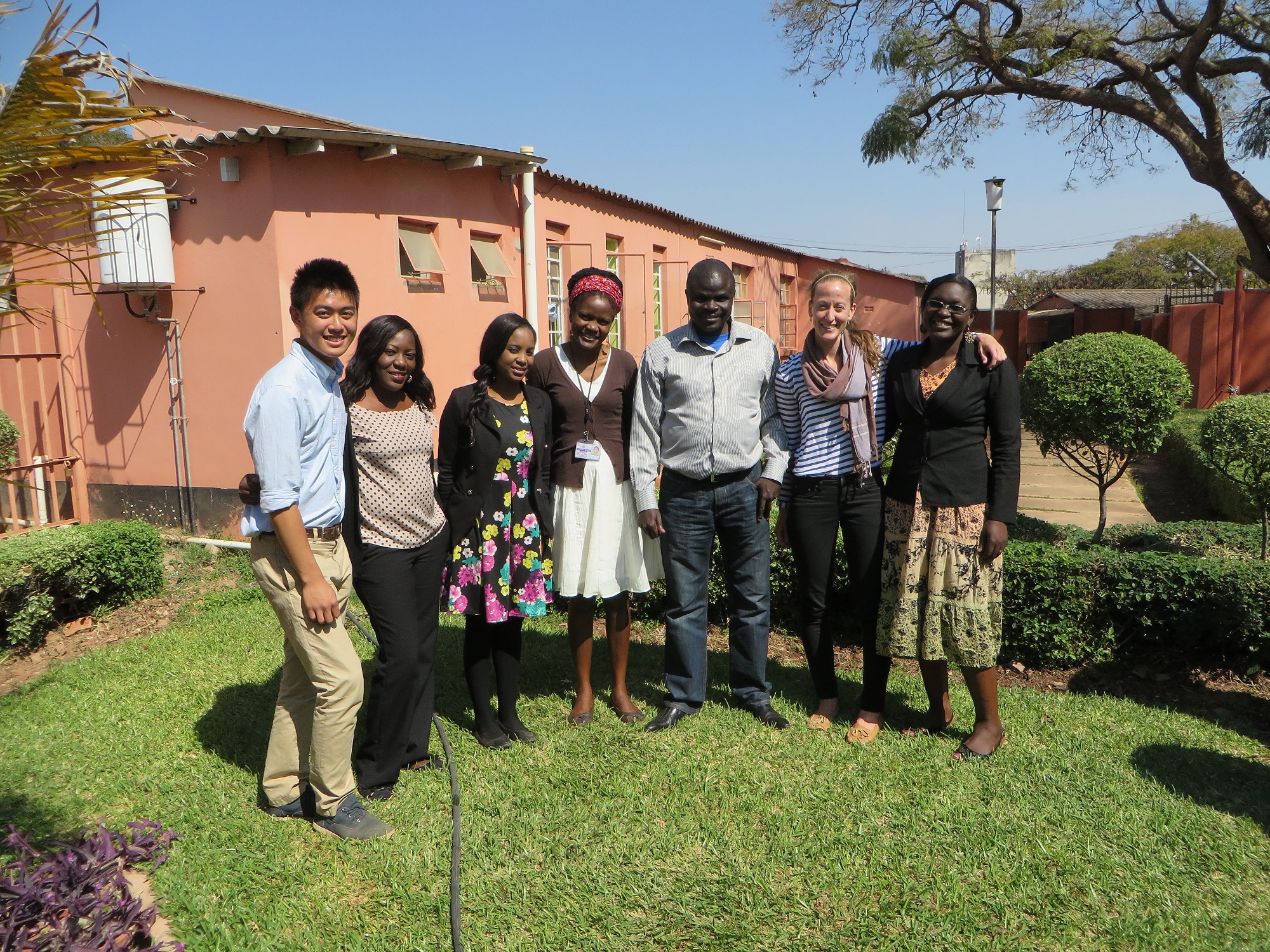
What’s it like to participate in a global health project abroad? MD student Jeffrey Wong spent this past summer working with expectant mothers in Zambia. He spoke to writer Suniya Kukaswadia about his experience.

Name: Jeffrey Wong
Program/year: Second year MD student
Role/position: Participant in the 2014 International Health Summer Research Program
Tell us about your work with the International Health Summer Research Program.
I went to Zambia this past summer to take part in a project involving pregnant women, some of whom were HIV-positive. The research was part of a longer-term initiative that looked at the effects of antiretroviral medications. I worked closely with nurses, midwives, physicians and staff from the University of Zambia to recruit women for the study, as well as design questionnaires and databases. I was also able to connect with the local Ministry of Health and clinic administrative staff to better understand the procedures necessary for initiating a research study in this setting. I keep in touch with the team in Zambia who say the data collection is going really well.
What inspired you to go to Zambia?
It was the perfect opportunity for me to further explore my interest in global health, particularly in the areas of research and clinical work. I wanted to better understand how research is conducted in a developing nation and the dynamics of a long-term global health research partnership.
What did you find most exciting about your time there?
I really enjoyed shadowing health professionals in clinics and during surgeries at the academic hospital. The experience helped me understand what it’s like to do clinical work in a lower resource setting. I realized just how important history taking and physical examinations were in a place where costly diagnostic tools aren’t readily available. Sometimes medical students forget to appreciate certain clinical skills because we have diagnostic tools to help us rule in/out conditions. I learned a lot of useful skills from the Zambian physicians.
I also got to discover Zambia’s beautiful culture. I really enjoyed exploring the vibrant city of Lusaka and bartering in my limited Bemba and Nyanja — Zambia’s two main languages. The Lusaka National Museum and annual agricultural fairs were a lot of fun. I was even able to visit Victoria Falls to experience “the smoke that thunders,” one of the seven natural wonders of the world.
What did you hope to accomplish during your time in Zambia?
I wanted to assist in a project that really changed patients’ lives. Women who participated in previous stages of the project would drop by the clinic to show their gratitude for the team’s help—it was really rewarding. Past research had helped reduce mother-to-baby HIV transmission rates.
I also wanted to see how I could incorporate global health research into my future career. The experience helped guide my focus towards global health. I plan on participating in global health electives during my clerkship so I can better appreciate a clinician’s role in low-resource settings.
What's your favourite thing about the Faculty of Medicine?
There are so many opportunities here. Whether you’re interested in a rural clinical experience, cutting-edge basic science research or global health opportunities, the Faculty has resources to achieve your goals. I wouldn’t have been able to go to Zambia if it weren’t for the International Health Summer Research Program.
Photo: University of Toronto students (Jeffrey Wong and Ashley Raeside) with Office Staff at the Department of Pediatrics at the University Teaching Hospital
Learn what are students are up to inside and outside of the classroom with Faces of U of T Medicine. From advising political leaders to providing care to Toronto’s most vulnerable populations, our students are making an impact on communities at home and around the world.
Do you have an interesting story to share? Contact Suniya Kukaswadia at suniya.kukaswadia@utoronto.ca
What’s it like to participate in a global health project abroad? MD student Jeffrey Wong spent this past summer working with expectant mothers in Zambia. He spoke to writer Suniya Kukaswadia about his experience.
Name: Jeffrey Wong
Program/year: Second year MD student
Role/position: Participant in the 2014 International Health Summer Research Program
Tell us about your work with the International Health Summer Research Program.
I went to Zambia this past summer to take part in a project involving pregnant women, some of whom were HIV-positive. The research was part of a longer-term initiative that looked at the effects of antiretroviral medications. I worked closely with nurses, midwives, physicians and staff from the University of Zambia to recruit women for the study, as well as design questionnaires and databases. I was also able to connect with the local Ministry of Health and clinic administrative staff to better understand the procedures necessary for initiating a research study in this setting. I keep in touch with the team in Zambia who say the data collection is going really well.
What inspired you to go to Zambia?
It was the perfect opportunity for me to further explore my interest in global health, particularly in the areas of research and clinical work. I wanted to better understand how research is conducted in a developing nation and the dynamics of a long-term global health research partnership.
What did you find most exciting about your time there?
I really enjoyed shadowing health professionals in clinics and during surgeries at the academic hospital. The experience helped me understand what it’s like to do clinical work in a lower resource setting. I realized just how important history taking and physical examinations were in a place where costly diagnostic tools aren’t readily available. Sometimes medical students forget to appreciate certain clinical skills because we have diagnostic tools to help us rule in/out conditions. I learned a lot of useful skills from the Zambian physicians.
I also got to discover Zambia’s beautiful culture. I really enjoyed exploring the vibrant city of Lusaka and bartering in my limited Bemba and Nyanja — Zambia’s two main languages. The Lusaka National Museum and annual agricultural fairs were a lot of fun. I was even able to visit Victoria Falls to experience “the smoke that thunders,” one of the seven natural wonders of the world.
What did you hope to accomplish during your time in Zambia?
I wanted to assist in a project that really changed patients’ lives. Women who participated in previous stages of the project would drop by the clinic to show their gratitude for the team’s help—it was really rewarding. Past research had helped reduce mother-to-baby HIV transmission rates.
I also wanted to see how I could incorporate global health research into my future career. The experience helped guide my focus towards global health. I plan on participating in global health electives during my clerkship so I can better appreciate a clinician’s role in low-resource settings.
What's your favourite thing about the Faculty of Medicine?
There are so many opportunities here. Whether you’re interested in a rural clinical experience, cutting-edge basic science research or global health opportunities, the Faculty has resources to achieve your goals. I wouldn’t have been able to go to Zambia if it weren’t for the International Health Summer Research Program.
Photo: University of Toronto students (Jeffrey Wong and Ashley Raeside) with Office Staff at the Department of Pediatrics at the University Teaching Hospital
Learn what are students are up to inside and outside of the classroom with Faces of U of T Medicine. From advising political leaders to providing care to Toronto’s most vulnerable populations, our students are making an impact on communities at home and around the world.
Do you have an interesting story to share? Contact Suniya Kukaswadia at suniya.kukaswadia@utoronto.ca
Optimize this page for search engines by customizing the Meta Title and Meta Description fields.
Use the Google Search Result Preview Tool to test different content ideas.
Select a Meta Image to tell a social media platform what image to use when sharing.
If blank, different social platforms like LinkedIn will randomly select an image on the page to appear on shared posts.
Posts with images generally perform better on social media so it is worth selecting an engaging image.
Suniya Kukaswadia
Prostate Cancer Researchers Develop Personalized Genetic Test to Accurately Predict Recurrence Risk
U of T researchers have developed a genetic test to identify which men are at highest risk for their prostate cancer to come back after localized treatment with surgery or radiotherapy.
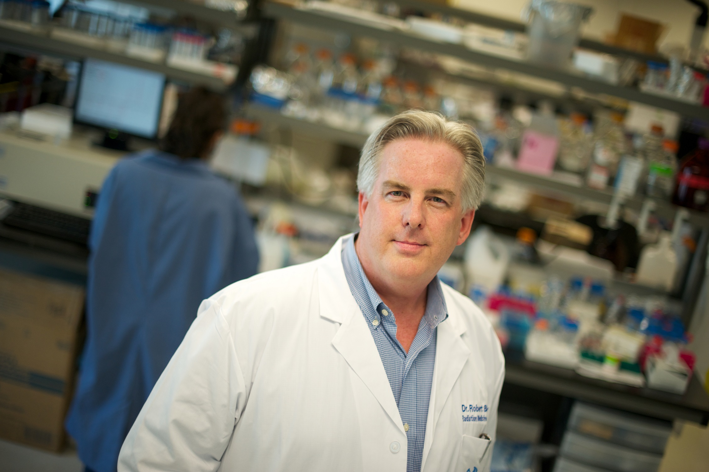
The findings are published online in Lancet Oncology. Professor Robert Bristow of the departments of Radiation Oncology and Medical Biophysics, and Assistant Professor Paul Boutros, of Medical Biophysics, report that the gene test provides a much-needed quick and accurate tool to determine with greater precision the men who will do well with local treatment only (surgery or radiation), and those who will need extra treatment (chemotherapy and hormone therapy) to ensure the cancer is completely eradicated. Bristow talks about the research here.
“Our findings set the stage to tackle the ongoing clinical problem of under-treating men with aggressive disease that will recur in 30 to 50 per cent of patients due to hidden, microscopic disease that is already outside the prostate gland during initial treatment,” says Bristow, a clinician-scientist at Princess Margaret Cancer Centre.
“This genetic test could increase cure rates in intermediate- to high-risk men by preventing progression to this metastatic spread of prostate cancer.”
The next step will be testing the gene signature on many more patients worldwide for three to five years to turn the test into one that is readily available in the clinic to guide personalized prostate cancer treatments.
The predictive test analyses biopsy tissue taken before treatment even starts to identify abnormal genetic characteristics (abnormal DNA) of the prostate cancer and its oxygen content. Low oxygen, or hypoxia, is an already known factor in the spread of prostate cancer. Together, this information can predict with almost 80 per cent accuracy – and in about three days – those prostate cancer patients who are at greatest risk of their disease returning, the study shows.
“The clinical potential is enormous for thousands of patients,” says Bristow. “This is personalized cancer medicine to the hilt – the ability to provide more targeted treatment to patients based on their unique cancer genetic fingerprint plus what’s going on in the cancer cell’s surrounding environment. We hope to improve cure rates by reducing the chances of the cancer recurring and prevent the cells from spreading.”
The researchers developed the genetic test with two groups of patients. In the first group, the team analyzed DNA from initial diagnostic biopsies of 126 men who were treated with image-guided radiotherapy (IGRT) and followed for an average 7.8 years. In the second group, the team used the test on 150 men whose tumours were removed surgically (radical prostatectomy). The genetic test produced similar results in both groups and therefore can be used in patients who choose radiotherapy or surgery as their initial treatment.
The researchers further found that when testing tumours for hypoxia in the men treated with IGRT and the gene test, this combined information made the test even more accurate, says Bristow.
The study showed that the men with the best outcomes – lower than seven per cent recurrence of prostate cancer at five years – had low levels of genetic changes and low hypoxia. For men with high levels of genetic changes and high hypoxia, outcomes were worse – more than 50 per cent of patients had recurrence and these are men who, in the future, could be offered intensified treatment as part of a personalized treatment plan.
Bristow and Boutros also co-lead the Canadian Prostate Cancer Genome Network (CPC-GENE) Sequencing Project.
Funding was provided by Prostate Cancer Canada, the Movember Foundation, the Ontario Institute for Cancer Research, and The Princess Margaret Cancer Foundation. The study was conducted with assistance from the Wellcome Trust Clinical Research Facility, Addenbrooke’s Clinical Research Centre, Cambridge, U.K.
Optimize this page for search engines by customizing the Meta Title and Meta Description fields.
Use the Google Search Result Preview Tool to test different content ideas.
Select a Meta Image to tell a social media platform what image to use when sharing.
If blank, different social platforms like LinkedIn will randomly select an image on the page to appear on shared posts.
Posts with images generally perform better on social media so it is worth selecting an engaging image.
Anxiety Can Damage Brain and Accelerate Conversion to Alzheimer’s
People with mild cognitive impairment (MCI) are at increased risk of converting to Alzheimer’s disease within a few years, but a new study warns the risk increases significantly if they suffer from anxiety.
The study, led by researchers at the University of Toronto and Baycrest Health Sciences’ Rotman Research Institute, has shown clearly for the first time that anxiety symptoms in individuals diagnosed with MCI increase the risk of a speedier decline in cognitive functions — independent of depression (another risk marker). For MCI patients with mild, moderate or severe anxiety, Alzheimer’s risk increased by 33 per cent, 78 per cent and 135 per cent respectively.
The research team also found that MCI patients who had reported anxiety symptoms at any time over the follow-up period had greater rates of atrophy in the medial temporal lobe regions of the brain, which are essential for creating memories and which are implicated in Alzheimer’s.
Until now, anxiety as a potentially significant risk marker for Alzheimer’s in people diagnosed with MCI has never been isolated for a longitudinal study to gain a clearer picture of just how damaging anxiety symptoms can be on cognition and brain structure over a period of time. A growing body of literature has identified late-life depression as a significant risk marker for Alzheimer’s. Anxiety has historically been subsumed under the rubric of depression in psychiatry. Depression is routinely screened for in assessment and follow-up of memory clinic patients; anxiety is not routinely assessed.
“Our findings suggest that clinicians should routinely screen for anxiety in people who have memory problems because anxiety signals that these people are at greater risk for developing Alzheimer’s,” said Linda Mah, principal investigator on the study, Assistant Professor in U of T’s Department of Psychiatry and Clinician-Scientist with Baycrest’s Rotman Research Institute. Dr. Mah is also a co-investigator in a multi-site study lead by the Centre for Addiction and Mental Health, and partially funded by federal dollars (Brain Canada), to prevent Alzheimer’s in people with late-life depression or MCI who are at high risk for developing the progressive brain disease.
“While there is no published evidence to demonstrate whether drug treatments used in psychiatry for treating anxiety would be helpful in managing anxiety symptoms in people with mild cognitive impairment or in reducing their risk of conversion to Alzheimer’s, we think that at the very least behavioural stress management programs could be recommended. In particular, there has been research on the use of mindfulness-based stress reduction in treating anxiety and other psychiatric symptoms in Alzheimer’s and this is showing promise,” said Mah.
The study accessed data from the large population-based Alzheimer’s Disease Neuroimaging Initiative to analyze anxiety, depression, cognitive and brain structural changes in 376 adults, aged 55 – 91, over a three-year period. Those changes were monitored every six months. All of the adults had a clinical diagnosis of amnestic MCI and a low score on the depression rating scale, indicating that anxiety symptoms were not part of clinical depression.
MCI is considered a risk marker for converting to Alzheimer’s disease within a few years. It is estimated that half-a-million Canadians aged 65-and-older have MCI, although many go undiagnosed. Not all MCI sufferers will convert to Alzheimer’s — some will stabilize and others may even improve in their cognitive powers.
The study has yielded important evidence that anxiety is a “predictive factor” of whether an individual with MCI will convert to Alzheimer’s or not, said Mah. Studies have shown that anxiety in MCI is associated with abnormal concentrations of plasma amyloid protein levels and T-tau proteins in cerebrospinal fluid, which are biomarkers of Alzheimer’s. Depression and chronic stress have also been linked to smaller hippocampal volume and increased risk of dementia.
In addition to Prof. Mah, the research team included Dr. Malcolm Binns (statistician scientist at Baycrest’s Rotman Research Institute, and Assistant Professor of the Dalla Lana School of Public Health at U of T), and Dr. David Steffens (Department of Psychiatry, University of Connecticut Health Centre).
The American Journal of Geriatric Psychiatry reported the findings online on October 29, ahead of print publication scheduled for May 2015.
The study was supported by the National Institutes of Health and the Geoffrey H. Wood Foundation.
Photo courtesy of Baycrest through iStock.
Optimize this page for search engines by customizing the Meta Title and Meta Description fields.
Use the Google Search Result Preview Tool to test different content ideas.
Select a Meta Image to tell a social media platform what image to use when sharing.
If blank, different social platforms like LinkedIn will randomly select an image on the page to appear on shared posts.
Posts with images generally perform better on social media so it is worth selecting an engaging image.

Dr. John McCrae, Toronto Medical Graduate

Last month, Dr. John McCrae was selected as a 2015 inductee to the Canadian Medical Hall of Fame. This timely announcement coincides with the centenary of the First World War, and the honour itself is a fitting commemoration of his famous poem, In Flanders Fields.
McCrae’s tribute to his fallen comrades, written on May 3, 1915 during a lull in the terrible battle of Ypres, was published in the London magazine Punch that December. It quickly became the most popular poem of the Great War, known throughout the British Empire and later in the United States. After a century the poppy and McCrae’s verses remain potent symbols of remembrance and the tragedy of war.
Yet the soldier-poet’s own story has been largely forgotten, except in his hometown of Guelph. There his memory is preserved in the limestone cottage where he was born in 1872, now a museum.
McCrae was a product of Ontario’s rural heartland. The grandson of rugged Scottish pioneers, he inherited his mother’s intellectual curiosity and warmth, his father’s love of the military, and his family’s strong Presbyterian commitment to duty. John and his brother Thomas (born in 1870) went on to become two of the University of Toronto’s most distinguished early medical graduates.
Even before entering U. of T., young John’s talent and broad interests were evident. He began writing poetry while at Guelph Collegiate, and at 14 joined the school’s cadet corps, winning a provincial gold medal. The following year he entered the militia unit commanded by his father. In 1888, at the age of 16, he became the first Guelph student awarded a University of Toronto scholarship.
McCrae spent most of the next decade in Toronto. Yet this key period in his life is sparsely documented, partly perhaps because the Faculty of Medicine (established in 1877) was still in its infancy. As an undergraduate at the university’s Knox (Presbyterian) College, he was a diligent student and devoted church-goer, but also found time to write poetry and remain active in both the Guelph militia and as one of the few members, along with writer Stephen Leacock, of the Varsity’s unit, “an honourable but neglected body called Company K.”
He excelled in biology under the supervision of Professor Ramsay Wright, earning the highest mark in his second year. In third year he slipped to second place as his chronic asthma worsened in the city smog. After taking a year off to teach at the agricultural college in Guelph, McCrae returned to Toronto in late 1893, completed his degree in Natural Sciences with high honours in 1894 and entered the Faculty of Medicine.
McCrae graduated in 1898, winning the gold medal as the top student in his class. Following a brief internship at the Toronto General Hospital, he joined the large contingent of top-notch Toronto medical graduates sent by Wright and J.E. Graham, professor of clinical medicine, to study with North America’s foremost medical educator. Canadian-born Dr. (later Sir) William Osler had transformed Johns Hopkins into the leading medical school and teaching hospital on the continent, and would go on in 1905 to become Regius Professor of Medicine at Oxford. McCrae’s brother Thomas, who had graduated from Toronto as silver medalist in 1895, remained at Hopkins as one of Osler’s assistant residents and eventually became one of his close colleagues.
McCrae spent several months working with Osler before beginning a fellowship in pathology at McGill University in September 1899. His studies were soon interrupted by the outbreak of the Boer War in mid-October. Believing that it was his duty to fight, he again took a year off, this time to lead the Guelph contingent of the Royal Canadian Artillery on active service in South Africa. There he served with distinction and was a popular officer, but was shocked by the cruelty and destruction of war.
After returning to Montreal in 1901, McCrae resumed his medical career, becoming one of the leading academic physicians of his generation, with positions at McGill University and the various teaching hospitals in addition to a large private practice. Gregarious and outgoing, he was also a popular social and literary figure with a large circle of friends.
McCrae resigned from the artillery brigade in 1904 after reaching the rank of Major, and was not involved with the military again until the eruption of the Great War in August 1914. As part of the British Empire, Canada was automatically involved in the conflict, and within three weeks 45,000 Canadians had rushed to enlist, McCrae among them. As before his chief motivation was duty rather than a naïve rush to adventure: “I am going because I think every bachelor, especially if he has experience of war, ought to go,” he wrote to a friend. “I am really rather afraid, but more afraid to stay at home with my conscience.”
Even so, nothing could prepare him for the horror of trench warfare. Appointed as brigade-surgeon to the Canadian Field Artillery, he found himself posted to the trenches near the devastated city of Ypres in Belgium. On April 22, 1915, the Germans launched an unexpected attack, using deadly chlorine gas for the first time in the war. The Canadian forces held the line for 17 days, but at a terrible cost. Half of the brigade was killed, including one of McCrae’s closest friends, who fell the day before he wrote In Flanders Fields.
McCrae was gratified by the success of his poem and its use as a call to arms, but his creative fires were now largely spent. It was the second-last poem he would write. Soon afterward he was promoted to lieutenant-colonel in charge of medicine at the No. 3 Canadian General Hospital in France staffed by his friends and colleagues from McGill. Those who had known him in happier times were shocked by the worn and exhausted man he had become. The enormous number of wounded and the grim reports from subsequent battles affected him deeply. Relentlessly committed to duty, he insisted on living in a tent throughout the winter like his comrades at the front and had to be ordered into the heated officers’ huts as his health failed. On January 24, 1918, McCrae was appointed as consulting physician to the First British Army, the first Canadian so honoured. But he did not live to take up this post. Four days later he died of pneumonia and meningitis, and was buried with full military honours in Wimereux, France.
McCrae’s tragic belief that he “would have broken faith had he lived while so many had died” — seen as an example of his selfless devotion to duty by his grieving compatriots, but now recognized as an ominous symptom of despair born of post-traumatic stress — remains the final chapter in this story of the terrible cost of war.
(Photo: Karen Roe Poppies by Karen Roe via Flickr)
Optimize this page for search engines by customizing the Meta Title and Meta Description fields.
Use the Google Search Result Preview Tool to test different content ideas.
Select a Meta Image to tell a social media platform what image to use when sharing.
If blank, different social platforms like LinkedIn will randomly select an image on the page to appear on shared posts.
Posts with images generally perform better on social media so it is worth selecting an engaging image.
Professor Edward Shorter and Sue Bélanger
